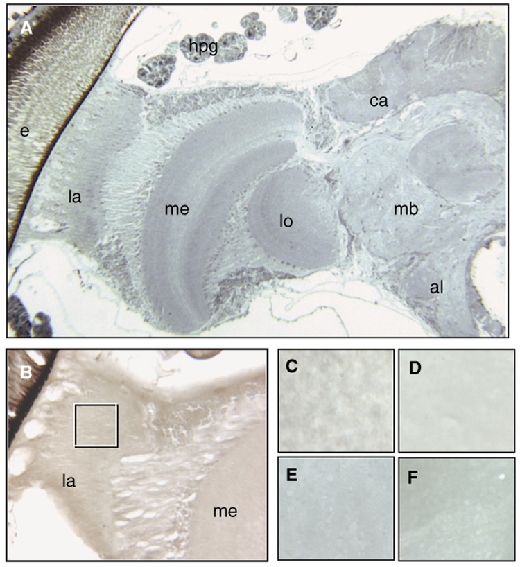
Fig. 1.

Overview of one-half of a honey bee brain (A) at 5×. Immunolocalization of nitrated proteins in cryosections (B–F) did not detect positive signals for nitration damage. The inserted box in (B) (16×, diutinus worker) refers to the area depicted in (C–F) (40×). (C) Nurse bee, (D) diutinus worker, (E) post-wintering forager, and (F) forager collected in summer. The differences in color between the pictures are optical artifacts and not due to positive staining. Abbreviations in (A and B): e, compound eye; la, lamina; me, medulla; lo, lobula; al, antennal lobe; mb, mushroom bodies; ca, calyx; hpg, hypopharyngeal gland (brood-food-producing glands). The images are representative for the full sample set (n = 30–50 per experimental group).
