
Fig. 4.
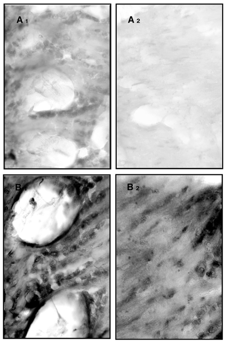
Immunolocalization of carbonylated proteins in cryosections (A and B). Close ups (40×) of lamina (1) and medulla (2) show that between-group differences in protein carbonylation are linked to social role rather than chronological age. The close up of 200-day-old nurse bees show some staining, mainly in the lamina (A1), with scattered staining in the medulla (A2), whereas 200-day-old foragers have high amounts of carbonylated proteins in both the lamina (B1) and medulla regions (B2).