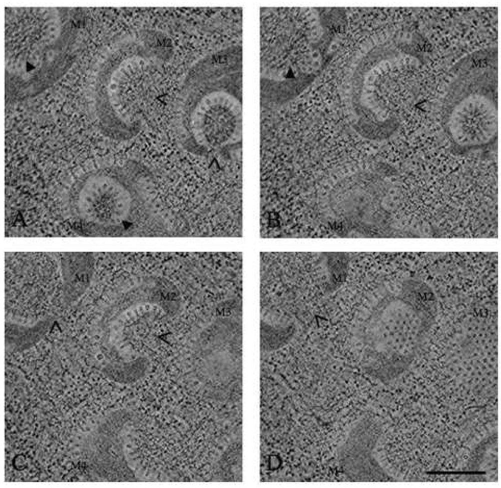
Fig. 4.
EMT of FHV in Drosophila cells. Drosophila cells at 12 h post infection were subjected to HPF and FS, embedded, and cut into ~500 nm sections. Two orthogonal −60 to +60 degree tilt series in 2 degree increments were collected. Four sections approximately 120 nm apart and about 12 nm thick were extracted from the reconstructed volume (A, B, C, and D). Four mitochondria were observed in all the sections and are labeled M1, M2, M3, and M4. A closed arrow indicates the sections where the mitochondrial chambers are closed to the cytoplasm. Open arrows indicate the openings in the mitochondrial chambers. Scale bar: 500 nm.

