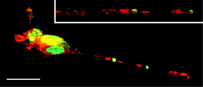
Figure 2.
Confocal microscopy image of NSC34/mAR cells expressing the mutant SBMA AR and treated with testosterone. Several SBMA AR neuropil aggregates have formed (green), along with an accumulation of the motor protein kinesin (red). Scale bar = 20 μm. Inset: closer view of a neurite. (After Piccioni et al, 2002.)

