
Figure 2. Analysis of pigments and chlorophyll precursors in the chlm mutant.
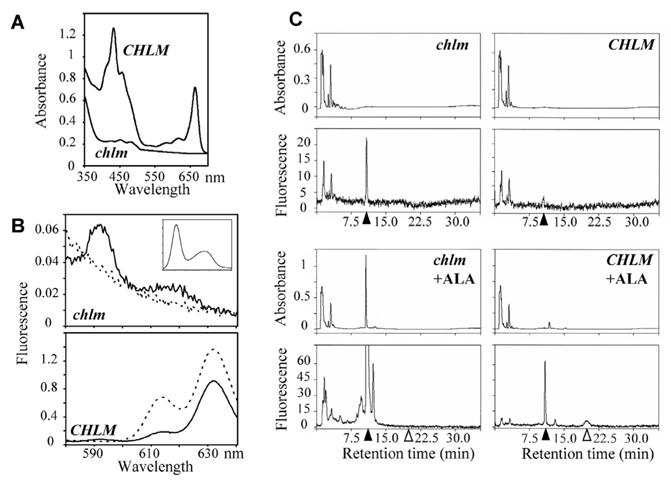
A- Absorbance spectra of pigments extracted from the mutant (solid line) and wild type/heterozygous plants (dotted line). B- Room temperature fluorescence emission spectra of acetone extracts of mutant and wild type/heterozygous plants. The plain line indicates emission obtained with excitation light at 402 nm and the broken line at 440 nm. Plants were grown for 10 d with a 10 h light cycle at 70 μmol photons m−2 s−1. The insert indicates fluorescence emission of standard Mg protoporphyrin IX with excitation light at 402 nm. C-HPLC traces of the acetone extracts of mutant and wild type/heterozygous plants. Plants were grown for 5 d at 70 μmol photon m−2 s−1, then for 15 d at 5 μmol photons m−2 s−1. 20 to 50 mg leaves + cotyledons were extracted. The eluate was monitored by absorbance at 420 nm (top) and by fluorescence emission at 590 nm with excitation set at 420 nm (bottom) for more specific detection of Mg protoporphyrin IX and Mg protoporphyrin IX methylester. Products were identified by concordance with retention times of standard compounds; closed and open arrows indicate position of Mg protoporphyrin IX and Mg protoporphyrin IX methylester, respectively. In +ALA conditions, plants were incubated overnight with 10 mM ALA and 5 mM MgCl2 in 10 mM Hepes pH 7.0 before extraction.