
FIGURE 2.
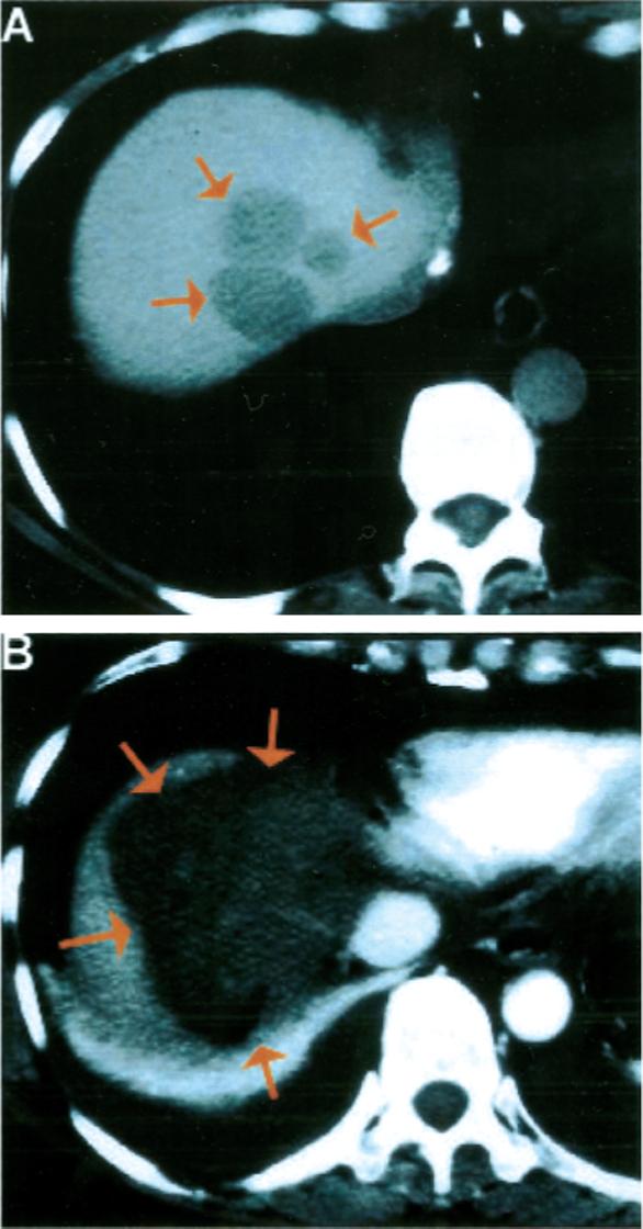
Colorectal carcinoma metastasis to the liver treated with open surgical radiofrequency ablation. (A) Contrast-enhanced computed tomography (CT) scan of the liver demonstrated three adjacent hepatic metastases (arrows) from colorectal carcinoma at the extreme dome of the liver in a difficult-to-treat location adjacent to the inferior vena cava and hepatic veins. (2) Contrast-enhanced CT scan of the liver on the day after treatment demonstrated the thermal lesion encompassing the region of liver containing the metastases, suggesting complete treatment. A pleural effusion also was noted, caused by the proximity of the diaphragm to the thermal lesion.