
FIGURE 3.
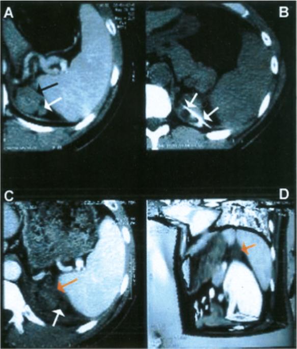
Adrenocortical carcinoma recurrence treated with percutaneous radiofrequency ablation. (A) Contrast-enhanced computed tomography (CT) scan demonstrated enhancing bilobed tumor (arrow) in adrenal bed. (B) CT scan during treatment demonstrated ablation needle (arrows) in tumor. (C) Enhanced CT scan after treatment depicted a lack of enhancement in tumors (arrows) and sliver of adjacent spleen, consistent with coagulative necrosis and cell death. (D) Three-dimensional enhanced CT image with planes cut away demonstrated treated tumor (arrow), with close proximity of adjacent nontarget organs (spleen, pancreas, kidney, stomach).