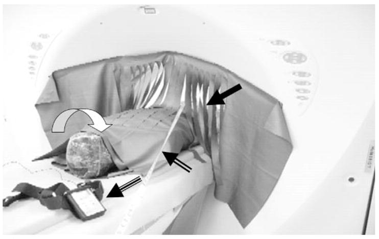
Figure 1.

The fenestrated curtain drape (closed arrow) hangs from the CT gantry. The phantom is wrapped in a 360° double layer of shielding. The tape measure (two-line arrow) is used to determine the distances from the scan plane. The curved white arrow indicates the phantom head; the three-line arrowhead indicates the dosimeter.
