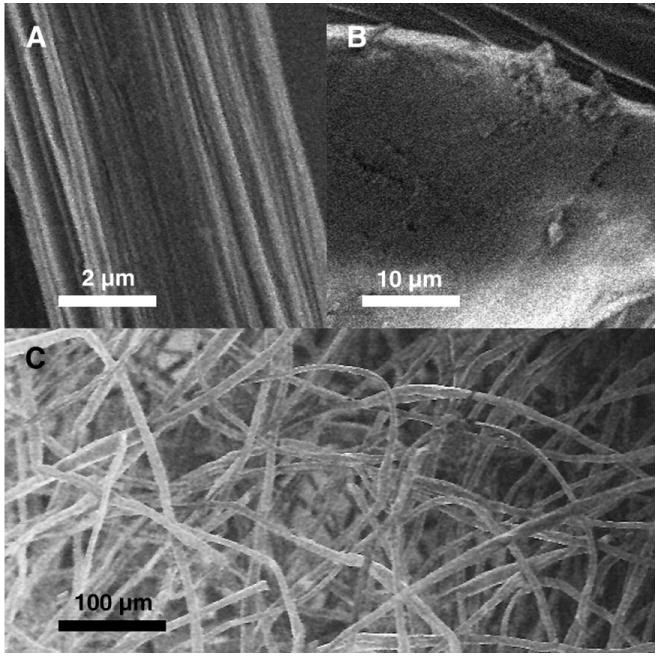
FIG. 1.

Scanning electron micrographs of the single PEC fiber as well as a needle-punched nonwoven fibrous scaffold. (A) Core fiber segment with striations on the surface. (B) Bead region with a smoothed surface. (C) Macroscopic view of nonwoven fibrous scaffold possessing high porosity.
