
FIG. 3.
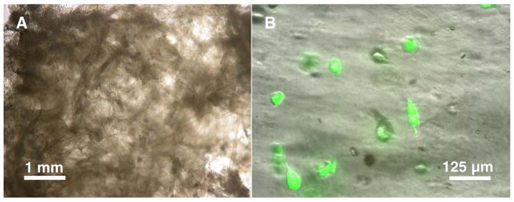
Images of blank and cell-seeded scaffolds. (A) When wetted, scaffolds swelled visibly and decreased in opacity but still retained their fibrous morphology, with most of the swelling confined to the bead regions. (B) Cells seeded on the scaffold remained viable even after 3 weeks of culture, although some cells had detached from the fibers, resulting in decreased cell seeding density.