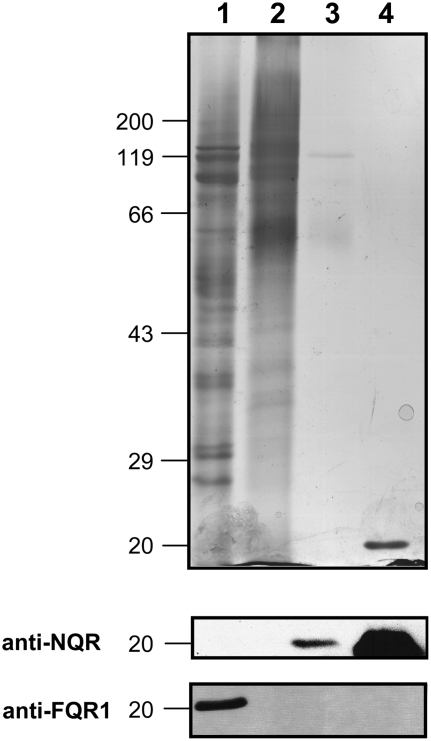
Figure 8.
Silver-stained SDS-PAGE (8%) and immunoblot analysis of protein fractions containing cytosolic proteins (lane 1), solubilized plasma membrane proteins (lane 2), purified NAD(P)H oxidoreductase protein (lane 3), and recombinant AtNQR (1 μg; lane 4). Samples with the following activities were loaded onto the gel for silver staining: 8 nmol XTTH2 min−1 (15 μg; lanes 1 and 2) and 140 nmol XTTH2 min−1 (7.5 μg; lane 3); samples with the following activities were loaded onto the gel for blotting: 30 nmol XTTH2 min−1 (lanes 1 and 2) and 140 nmol XTTH2 min−1 (lane 3). Marker positions (kilodaltons) are given on the left side. Immunoblots were decorated with antibodies directed against NQR and FQR1. The amount of NQR protein in the plasma membrane protein fraction (lane 2) was insufficient for detectable NQR antibody binding.

