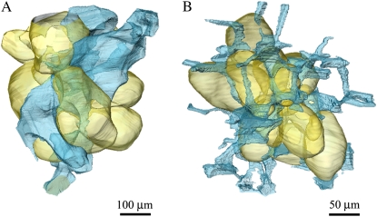
Figure 2.
A and B, 3-D rendering of parenchyma tissue and single cells (in yellow) of apple (A) and pear (B) with adjacent voids (in blue). Images obtained from phase contrast tomography. While the voids lie like wires of a tight net around the pear cell, a small number of larger voids connect in an irregular disconnected pattern to the apple cell.

