
Figure 7.
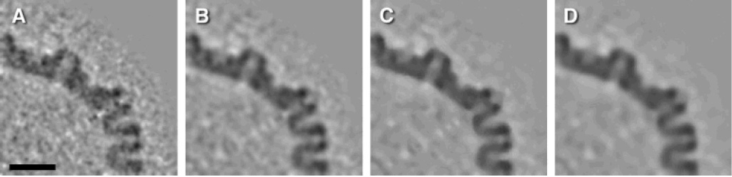
Noise reduction using different denoising methods. One HSV-1 capsid was extracted from a tomogram and 60-fold symmetrized. (A) Central section of capsid after symmetrization. (B–D) Central section of capsid after symmetrization and denoising with different filters: (B) bilateral filter, (C) median filter (three iterations), (D) anisotropic non-linear diffusion (60 iterations). Bar: 50 nm.