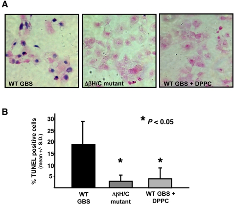
Figure 2. GBS βh/c effects on HL-1 cardiomyocyte viability.
(A) Trypan blue staining of HL-1 cells exposed for 1 h to extracts (1∶200 dilution) from WT GBS in the absence or presence of DPPC (2 mg/ml) or to similar extracts of Δβh/c mutant GBS. (B) Analysis of TUNEL staining by flow cytometry. Percent TUNEL positive HL-1 cells that had been treated for 1 h with extracts (1∶200) of WT GBS in the absence or presence of 2 mg/ml DPPC or with extracts from Δβh/c mutant GBS. Apoptosis data represent the mean +/− SD from three separate experiments.

