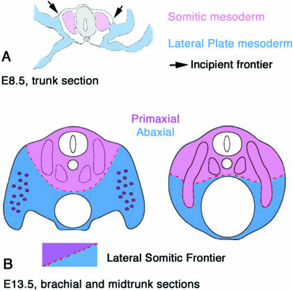
Fig. 1.
Schematic illustrations of the vertebrate mesoderm and the definitions and dispositions of the primaxial and abaxial domains separated by the lateral somitic frontier. (A) Cross-section of the midtrunk region of an embryonic day 9 mouse embryo. The paraxial somites (so) are pink, somatic LP mesoderm is blue (lp). For simplicity the intermediate mesoderm is not defined. (B) Cross-section at approximately embryonic day 11. Primaxial domain is pink, abaxial domain is blue, and includes somitic myoblasts that are illustrated in purple.

