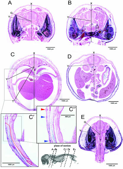
Fig. 3.
A series of transverse sections along the A–P axis of E15.5 Prx1cre:Z/AP embryos. Lines are used to illustrate differences in the expansion of the body wall and variation in the cross-sectional profile of the label boundary. Note the variation in topography at different levels. ‘X’ marks the dorsal point of the saggital midline. ‘Y’ marks the superficial label boundary, which approximates to the original boundary between LP and somitic mesoderm. Angle ‘XY’ marks the extent of angular displacement of the label boundary from the saggital midline. In (A,E) the labelled domain is expanded around the limb girdles (points ‘G’) dorsal to the superficial label boundary. (A) Brachial level; the dorsalmost extent of the labelled domain in the pectoral girdle is marked by point ‘G’. (B) Brachial-to-thoracic transition. (C) Thoracic level; the unlabelled thoracic wall comprising ribs and intercostal muscles has displaced the frontier to point ‘T’. (C′) Higher magnifications of the flank region in (C) with the label boundary marked with a red dashed line. (C″) Further magnification of the area of the boundary. Note the three hair follicles, two developing in the labelled domain (blue arrowhead) and one developing in the unlabelled domain (red arrowhead). (D) Lumbar region. (E) Sacral level; the dorsalmost extent of the labelled domain in the pelvic girdle is marked by point ‘G’.

