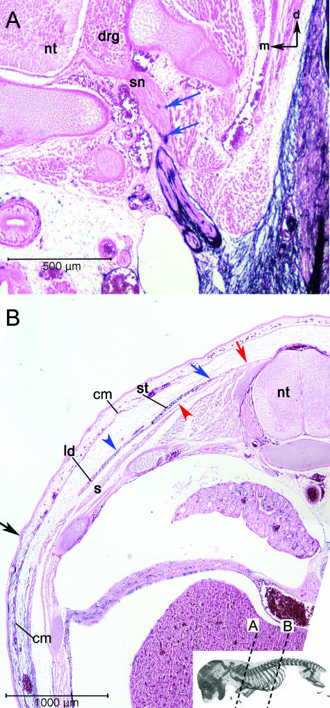
Fig. 5.
Extreme discontinuities in the label boundary. (A) AP-labelled cells (blue arrows) are associated with spinal nerves (sn) of the brachial plexus close to the point where they emerge from the vertebral column. This is more proximal than the rest of the label boundary. Black arrows in upper right orientate dorsal (d) and medial (m). (B) The dorsolateral superficial muscles of the brachial/thoracic region also cause discontinuities in the label boundary. The cutaneous maxiums (cm) is unlabelled at its insertion on the subcutaneous fascia of the dorsal trunk although it is labelled at its origin on the humerus. The frontier in the cm is continuous with the label boundary in the surrounding mesenchyme (black arrow = superficial label boundary). The blue arrowhead and arrow indicate the labelled region the spinotrapezius (st) and the latissimus dorsi (ld), respectively. These muscles are unlabelled at their origins on the vertebrae and thoracolumbar fascia (red arrow and arrowhead) and labelled at their insertion on the scapula and humerus, respectively. The label boundary in these mucles is far removed from the major par of the boundary, which angles ventrally through the body wall from its superficial boundary (black arrow). The red arrowhead indicates the unlabelled region of the muscle. The underlying serratus dorsalis (s) contains no blue label. Abbreviations: neural tube (nt); spinal nerves (sn); spinotrapezius (st); latissimus dorsi (ld); serratus dorsalis (s).

