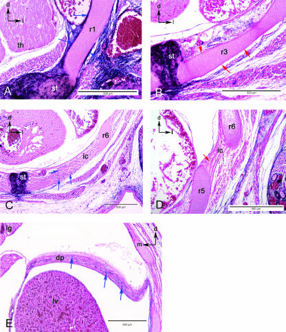
Fig. 6.
Sections in the thoracic region. (A) The chondrocytes of the first rib (r1) are somite derived, yet the periosteum is labelled (blue arrows) as is the associated connective tissue and the sternum (st). Black arrows in upper left indicate dorsal (d) and lateral (l). (B) View of the third rib (r3), where the periosteum is unlabelled (red arrows) although the surrounding connective tissue is labelled blue. Black arrows in upper left indicate dorsal (d) and lateral (l). (C) View of the intercostals muscle (ic) at the level of the sixth rib (r6). Distal portion of the intercostal muscles are associated with blue-labelled cells (blue arrows). (D) Proximal portion of the intercostal muscles (ic) do not include blue cells (red arrow). (E). Section through the diaphram (d) showing the inclusion of blue-labelled cells (blue arrows). Black arrows in upper corners orientate dorsal (d), lateral (l) or medial (m). Abbreviations: intercostals muscle (ic); liver (lv); lung (lg); thymic rudiment (th).

