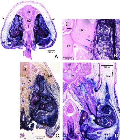
Fig. 9.
General view of the sacral level. (A) The bulk of the pelvic girdle appears to dorsally expand the labelled domain above the superficial label boundary (black arrows). (B) Higher magnification of a sacral rib (sr). AP-positive cells, apparently chondroblasts (blue arrows), are included in the cartilage at the distal end of the sacral rib where it meets the ilium (il). (C) The bones of the pelvis are labelled; the gluteus maximus muscle (gm) is associated with labelled connective tissue as it inserts on the proximal femur and at its origin on the ilium, although it is associated with unlabelled connective tissue at its origin from the lumbar fascia. (D) Insertion of the psoas major (pm) and iliacus (ilc) onto the lesser trochanter of the femur (f). Black arrows in upper right corner orientates dorsal (d) and medial (m). Abbreviations: lumbar vertebrae (lv); neural tube (nt); sacral vertebrae (sv); sacral rib (sr); ilium (il); femur (f); ischium (is); psoas major (pm); iliacus (ilc).

