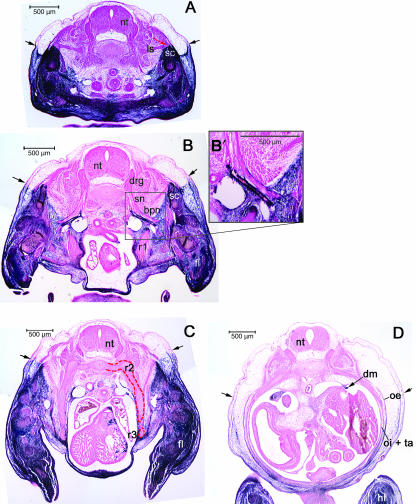
Fig. 10.
The label boundary in E13.5 mice. (A) At prechondrogenic stages the scapula (sc) has both labelled and unlabelled (red arrow) domains. The unlabelled levator scapulae (ls) can be seen inserting upon the vertebral border (red arrow). The labelled domain rises above the superficial label boundary (black arrows). (B) At deeper brachial levels, the distal tip of the pre-cartilage condensation of the first rib (r1) is completely surrounded by AP-stained mesenchyme. (B′) Higher magnification view of the subclavian artery (sbc) and the nerves of the brachial plexus (bp) that cause a partial discontinuitity in the label boundary while entering the brachial region. (C) Thoracic level on E13.5 showing the unlabelled expansion containing the second and third ribs (r2 and r3, red outline) with labelled intercostals muscle between them (blue arrow) ventrally displacing the label boundary from its superficial location (black arrows). (D) At the lumbar level the obliquus externus (oe), obliquus internus (oi) and transversus abdominis (ta) show staining that increases in intensity in the dorsal-to-ventral direction. The dorsal attachment of the diaphram (dm) to the coelom wall includes many blue cells. Abbreviations: hindlimb (hl); scapula (sc); neural tube (nt); dorsal root ganglion (drg); spinal nerve (sn); forelimb (fl).

