Fig. 1.
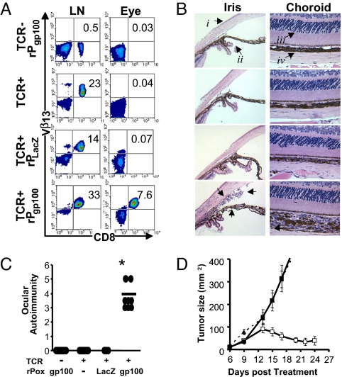
Induction of ocular autoimmunity during effective tumor destruction. (A) Flow cytometric analysis of Vβ13+CD8+ MDA-reactive pmel-1 T cells in the inguinal lymph node or eye 5 days after exogenous IL-2 and recombinant poxviral immunization of pmel-1 (TCR+) or WT (TCR−) mice. (B) Eyes from A were H&E-stained and examined for changes in morphology of the cornea (i), iris (ii), photoreceptors (iii), or choroid (iv). Arrows highlight cellular infiltrates in pmel-1 mice receiving recombinant gp100 poxvirus and IL-2. Data are representative of two independently identically performed experiments. (C) Ocular autoimmunity at day 5 after IL-2 and vaccination were assessed by using a masked ocular autoimmunity score as described in Methods from two independently performed experiments, *, P > 0.0001 vs. pmel-1 without vaccination. (D) Treatment of established tumors in pmel-1 transgenics vaccinated with exogenous IL-2 and recombinant gp100 poxvirus (□), without vaccination (■), or vaccinated WT littermates (▴), representative of two independent experiments.

