
Figure 3. Expression of BMP4 in the fetal liver of f/f and control mice.
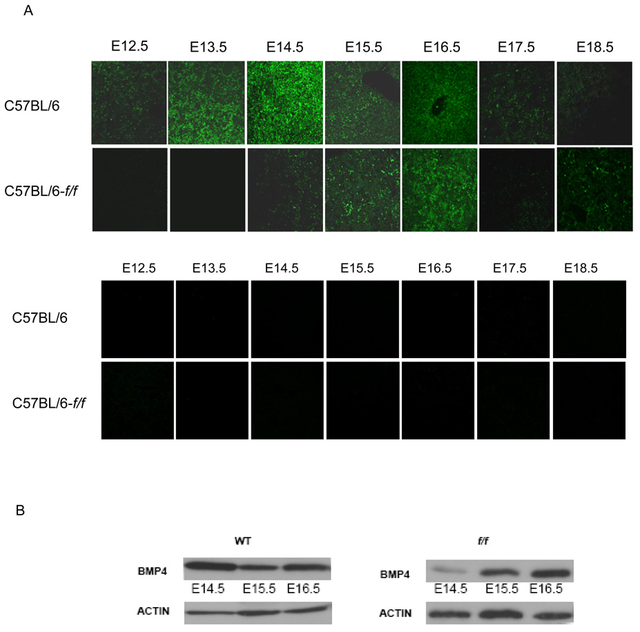
(A) (top) Fetal livers from control and f/f embryos isolated at the indicated days were sectioned and stained with anti-BMP4 antibodies. (Bottom) Negative control staining with isotype control antibodies of fetal liver sections from C57BL/6-f/f and C57BL/6 control embryos. (B) Western blots of whole fetal liver lysates from the indicated days of development probed with anti-BMP4 antibodies and anti-actin antibodies as a loading control.