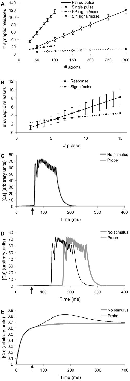
Figure 2. Designing the Stimulus Protocol.
(A) Baseline stimulus distribution scaling with number of axons. Paired pulse stimulus had better signal-to-noise and required fewer input axons than single pulse. (B) Probe stimulus scaling with number of pulses. Signal-to-noise (on same axis) improved slowly. (C, D, E) Comparisons of baseline (no stimulus) and baseline+probe responses at −65, −66 and −67 mV reversal potential for potassium. The curves were separable only at −66 mV and even at this potential the cells went into spontaneous bursts.

