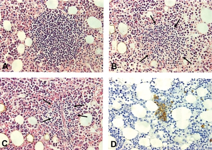
Figure 4.
Histopathological appearances in bone marrow trephine biopsy from a patient with primary CAD. Lymphoid infiltrates may be of variable size; large (A), medium-sized (B), or often small and poorly outlined (C) which renders them barely detectable within areas of hyperplastic erythropoiesis unless immunohistological staining is applied (D). A–C, HE-stain; D, Anti-CD20, horseradish peroxidase/diaminobenzidine. All photomicrographs are taken at identical magnification (× 40 objective) to enable comparison of individual infiltrates.

