
FIG. 2.
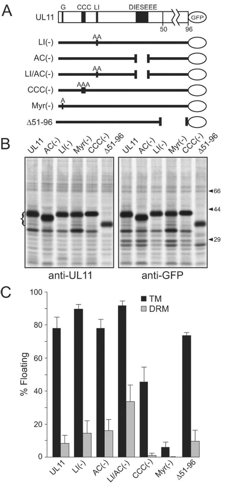
DRM association of UL11 mutants. (A) Diagram of UL11-GFP and the mutants that were analyzed. The motifs of interest are shown: G, myristylation site; CCC, palmitylation site; LI, di-leucine-like; DIESEEE, acidic cluster (AC). Sites of alanine substitutions are indicated. (B) To examine the reactivity of the mutants to anti-UL11 and anti-GFP sera, transfected cells were metabolically labeled and immunoprecipitated proteins were analyzed by SDS-PAGE. The positions of the UL11-GFP species are indicated with a bracket to the left. Positions of markers (in kDa) are indicated to the right. (C) Constructs depicted in panel A were analyzed for their ability to float in the absence (TM) and presence (DRM) of 0.5% TX-100. Each construct was analyzed a minimum of three times.