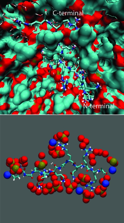
Fig. 1.
Binding of a Tat peptide to a zwitterionic lipid bilayer. (Upper) Snapshot obtained at the end of the 200-ns simulation (simulation A in Table 1). The peptide is partially exposed to water and surrounded by phosphates and carbonyl groups of the phospholipids. (Lower) Positively charged groups of the peptide bind to the phosphate and carbonyl groups of the phospholipid molecules. In this configuration, the Tat peptide binds to 14 phosphate groups.

