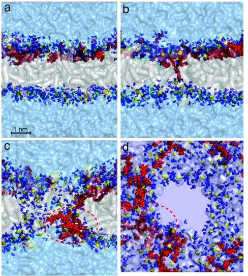
Fig. 5.
Snapshots at different times along the simulation of a system composed of 92 DOPC lipids, 8,795 water molecules, and four Tat peptides (simulation G in Table 1). Colors and representations are the same as in Fig. 2. (a) Position of the peptides before translocation. (b and c) Translocation of an arginine amino acid (b) toward the distal layer that nucleates the formation of a water-filled pore (c). (d) Snapshot at the same instant as c but from a direction perpendicular to the membrane, showing another perspective of the pore and translocating peptide.

