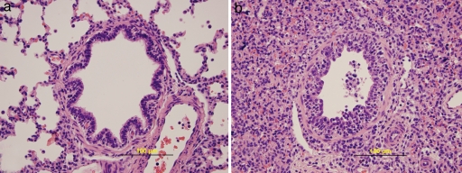
Fig. 2.
Microscopic lung sections from control and infected pigs. (a) Bronchiole in the lung of a control pig inoculated with noninfectious cell culture supernatant. Note the regular outline of the pseudostratified columnar epithelium. (b) Necrotizing bronchiolitis in the lung of a pig 3 days after inoculation with H2N3 swine influenza virus. The epithelial lining of the airway is focally disrupted by sloughing of necrotic infected cells and early reactive proliferation of the remaining epithelium. The lumen contains sloughed epithelial cells and mixed leukocytes. A small number of lymphocytes are seen infiltrating subepithelial and peribronchiolar connective tissue.

