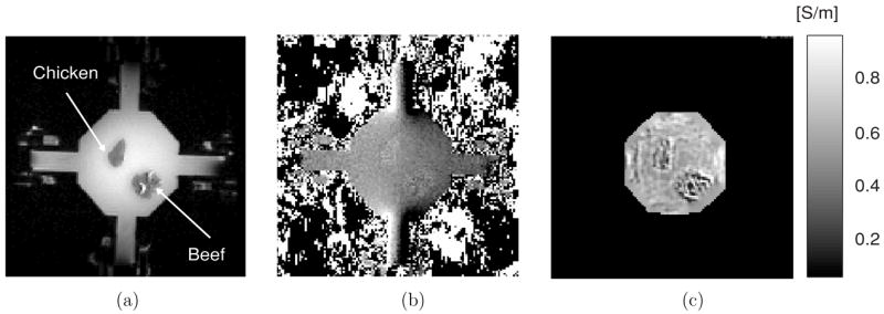
Figure 8.

MREIT experiment at 5 mA injection current. (a) MR magnitude image of a 40 mm tissue phantom containing pieces of chicken breast and bovine muscle. (b) Magnetic flux density image. (c) Reconstructed conductivity image. A SE pulse sequence was used with TR/TE = 600/10 ms, I = 5 mA and Tc = 9 ms. Eight slices were imaged with 1 mm slice thickness, NEX was 16, FOV was 80 × 80 mm2, image matrix size was 128 × 128 and pixel size was 0.625 × 0.625 mm2.
