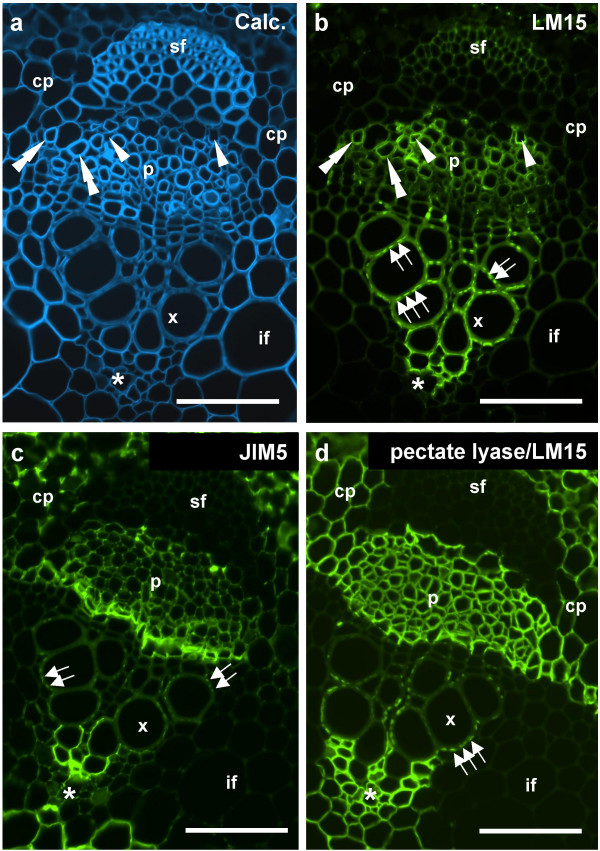
Figure 6.
Indirect immunofluorescence detection of xyloglucan and pectic HG epitopes in TS pea stem internode vascular bundles. a. Calcofluor White image of section showing all cell walls. b. LM15 binding to an equivalent section to a. The antibody binds most strongly to a region of protoxylem but also certain cells in the phloem region. c. JIM5 binding to an equivalent section shows binding to protoxylem and cambial cells. d. LM15 binding to an equivalent section pre-treated with pectate lyase shows the epitope detected abundantly in the phloem/cambial regions and cortical parenchyma. Arrowheads indicate cells in the phloem regions without thickened cell walls in which the LM15 epitope is detected without pre-treatment. Double arrowheads indicate cells with thickened cell walls/LM15 epitope in the phloem region. Sets of arrows indicate the punctuate presence of LM15 and JIM5 epitopes in xylem vessel cell walls. Asterisk indicates distal extent of the protoxylem. cp = cortical parenchyma, p = phloem region, pf = phloem fibre bundle, x = xylem vessel, if = interfascicular fibres. Scale = 10 μm.

