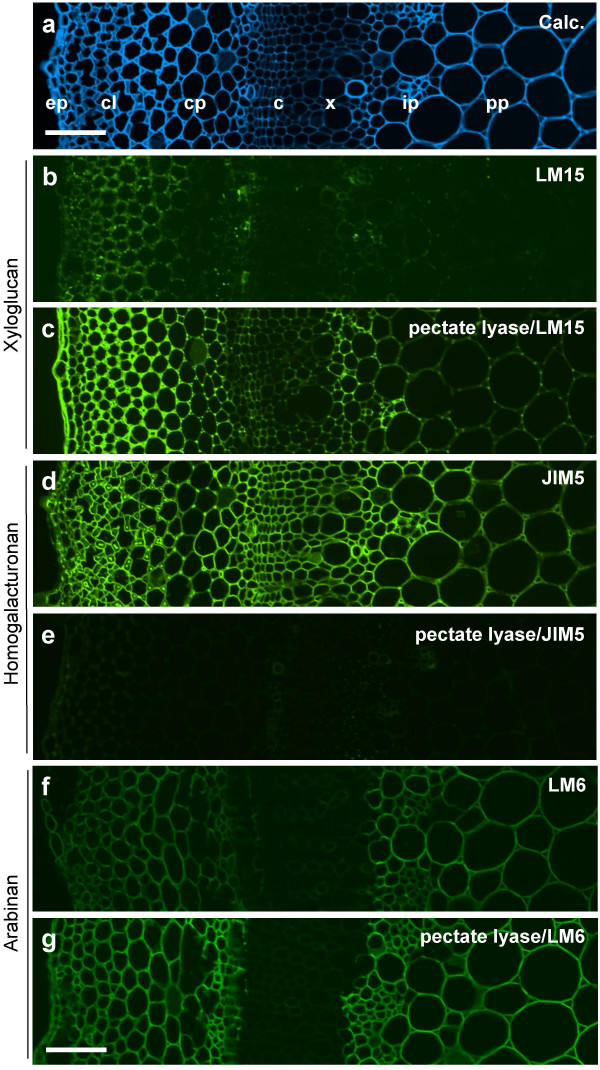
Figure 7.
Indirect immunofluorescence detection of xyloglucan, pectic HG and arabinan epitopes in TS tobacco stem internode. a. Calcofluor White image of section showing cell types from epidermis to pith parenchyma. b. LM15 binding to an equivalent section to a. There is weak recognition of cortical collenchyma/parenchyma and isolated groups of cells in the cambial region. c. An equivalent section to b that had been pre-treated with pectate lyase to remove pectic HG. LM15 binds strongly to epidermal and parenchyma cell walls. d. Section immunolabelled with pectic HG probe JIM5 which binds to all cell walls. e. Equivalent section to d pre-treated with pectate lyase indicates that the JIM5 epitope has been abolished. f. Section immunolabelled with arabinan probe LM6 which binds most strongly to cell walls of cortical and pith parenchyma. g. Equivalent section to f pre-treated with pectate lyase indicates increased detection of the same pattern of the LM6 epitope. ep = epidermis, cl = collenchyma, cp = cortical parenchyma, c = cambium, x = xylem, ip = internal phloem, pp = pith parenchyma. Scale = 100 μm.

