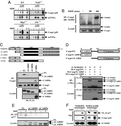
Fig. 2.
NOD2 binds to and activates caspase-1. (A) Caspase-1 activation by MDP is NOD2-dependent. Peritoneal macrophages from the indicated strains were treated as described in Fig. 1A, and supernatants were analyzed by immunoblotting for the presence of activated caspase-1 (p20) and TNF-α. L/A = macrophages were primed with LPS + ATP as in Fig. 1A. (B) NOD2 interacts with caspase-1 upon MDP stimulation. TDM were treated with MDP (10 μg/ml). At the indicated time points, cells were lysed, caspase-1 was immunoprecipitated (IP), and the presence of NOD2 in immunoprecipitates and original lysates was examined by immunoblotting (IB). (C) The NOD2 CARD motif binds caspase-1. Myc-tagged caspase-1 was coexpressed in HEK293T cells with indicated FLAG-tagged NOD2 constructs. After 36 h, cells were lysed and caspase-1 was immunoprecipitated. The presence of FLAG-tagged NOD2 and caspase-1 in the immunoprecipitates was examined by immunoblotting. (D) Caspase-1 binds NOD2 via its CARD motif. FLAG-tagged NOD2 was coexpressed in HEK293T cells with the indicated Myc-tagged caspase-1 constructs. After 36 h, cell lysates were immunoprecipitated with Myc antibody and analyzed as above. (E) The NOD2-CARD motifs are required for MDP-induced IL-1β secretion. FLAG-tagged NOD2 expression vectors were cotransfected into HEK293T cells along with caspase-1 and pro-IL-1β expression vectors as above. After 36 h, culture supernatants and cell lysates were examined for mature IL-1β and the different NOD2 proteins, respectively. (F) The NOD2-LRR prevents caspase-1 activation in the absence of MDP. The different NOD2 constructs were coexpressed in HEK293T cells as shown. Secretion of mature IL-1β and activated caspase-1 was examined as above.

