Fig. 5.
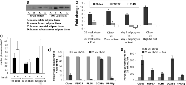
Rosiglitazone treatment of cultured adipocytes or intact mice markedly increases Cidea expression. (a) Western blot analysis of Cidea expression in mouse and human adipose tissues. (b) Fold change of Cidea, FSP27, and perilipin mRNA in 3T3-L1 adipocytes and primary adipocytes isolated from mice after rosiglitazone treatment based on expression data using MG-U74 Affymetrix GeneChips. Total RNA was isolated from 3T3-L1 adipocytes treated with or without 1 μM rosiglitazone for 24 h or primary fat cells of mice treated with or without 5 mg/kg rosiglitazone each day for 2 weeks. *, P < 0.05. (c) The graph represents amount of d-[U-14C]-glucose taken up and converted to triglycerides by primary adipocytes from 4-week-old ob/ob mice, 26-week-old ob/ob mice, and 26-week-old ob/ob mice treated with 5 mg/kg rosiglitazone each day for 2 weeks. Glucose conversion to triglyceride glycerol in adipocytes plus or minus insulin was calculated to nanomoles per 105 cells. The data represent the mean ± SEM of three experiments for each age group and condition. (d) Quantitative real-time analysis performed by using RNA isolated from adipose tissue (epididymal fat pads) of 4-week-old ob/ob and 26-week-old ob/ob mice. The 36B4 gene was used as a reference gene for quantitative analysis (P < 0.05). (e) Quantitative real-time analysis performed by using RNA isolated from adipose tissue (epididymal fat pads) of 26-week-old ob/ob mice and 26-week-old ob/ob mice treated with 5 mg/kg rosiglitazone each day for 2 weeks. The 36B4 gene was used as a reference gene for quantitative analysis (P < 0.05). All procedures in Fig. 5 were carried out according to the guidelines of the University of Massachusetts Medical School Institutional Animal Care and Use Committee.

