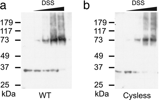
Fig. 1.

Hv1 is a dimer. Shown are Western blots of membranes from tsA201 cells transfected with WT (a) and Cysless (b) human Hv1 cDNAs and exposed to increasing concentrations of the amino group-specific cross-linker DSS (from left to right: no DSS, 25 μM, 75 μM, 250 μM, 2.5 mM). Membrane samples were run on SDS/PAGE under reducing conditions (see Methods). All of the gels that were used were 12%. Molecular mass markers are shown on the left.
