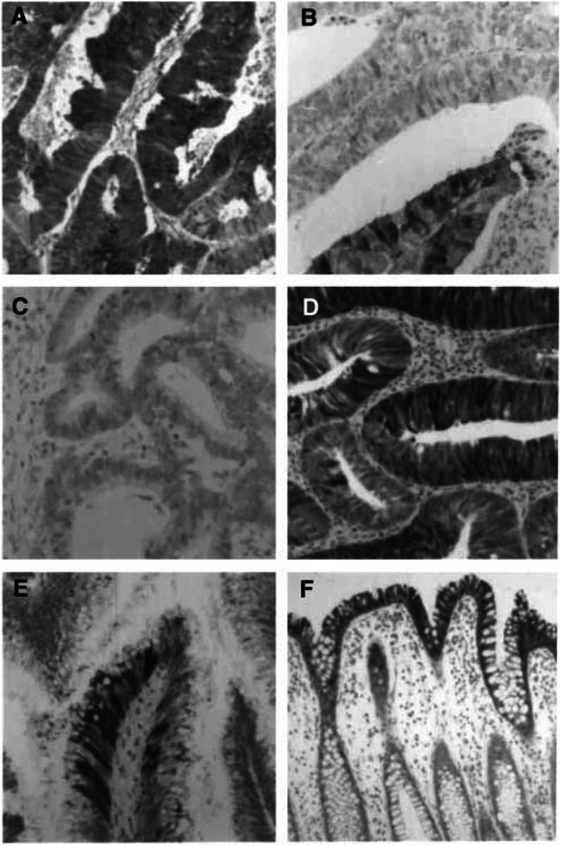
Figure 2.
The immunohistochemical localisation of L-FABP in colon cancer, colon adenomas and normal colon. In colon cancer there is patchy staining of the tumour cells. (A) Tumour with a high proportion of tumour cells staining positively for L-FABP while (B) is a tumour with a low proportion of L-FABP positive tumour cells. The patchy or mosaic staining for L-FABP is also demonstrated in (B). (C) Tumour that is negative for L-FABP. A small tubular adenoma (D) showing patchy staining for L-FABP. In a villous adenoma (E) there is only a small proportion of tumour cells showing L-FABP immunoreactivity and the staining is in discrete groups of cells. Immunohistochemical staining for L-FABP in normal colon is present in the surface epithelium and the upper half of the crypt epithelium (F).

