Figure 3.
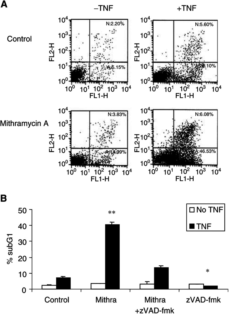
Sensitisation to TNF-induced apoptosis by mithramycin is caspase dependent. TF-1 cells were treated with 20 ng ml−1 TNF in the presence or absence of 75 nM mithramycin. (A) After 24 h of incubation, cells were stained with annexin V and PI. Necrosis (N) and apoptosis (A) were quantified by flow cytometry. The annexin V-positive cells are the cells undergoing apoptosis and are represented in the lower right quadrant. Similar results were obtained from two more independent experiments. (B) Effect of 50 μM caspase inhibitor zVAD-fmk. Percentage of subG1 cells was determined by PI staining and analysed by flow cytometry. *, P<0.05; **, P<0.01 of three independent experiments compared to TNF-treated cells.

