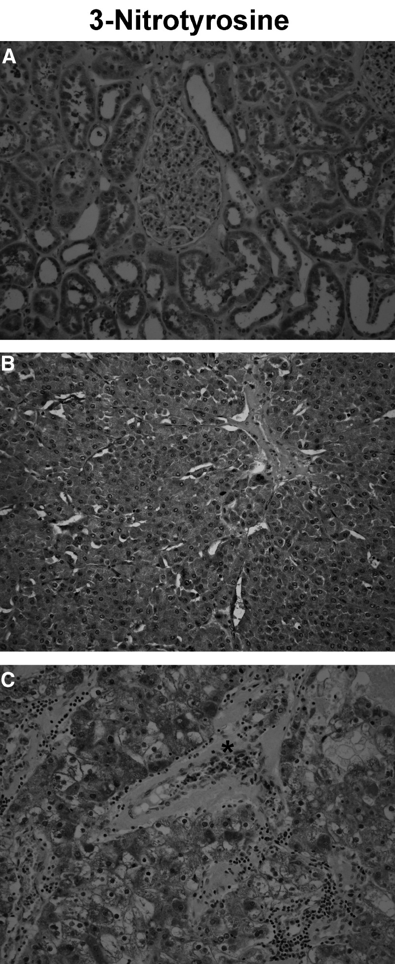
Figure 3.
Changes in the immunostaining pattern of protein tyrosine nitration between normal human kidney/benign tumour and RCC. Immunodetection of 3-nitrotyrosine on paraffin sections. (A) In normal renal parenchyma, most of the tubular epithelial cells along the nephron were labelled, with a variable staining intensity. In glomeruli, some of the podocytes and parietal cells scored positive. Red blood cells were not labelled. (B) In oncocytoma, a strong, granular and homogeneous staining of tumour cells was noted. Red blood cells were not labelled, and there was no inflammatory infiltrate. (C) In contrast, in malignant tumours (a case of grade III clear cell RCC is shown), the pattern of protein tyrosine nitration was heterogeneous. Red blood cells (*) were strongly stained and an inflammatory infiltrate was present (original magnification × 200).

