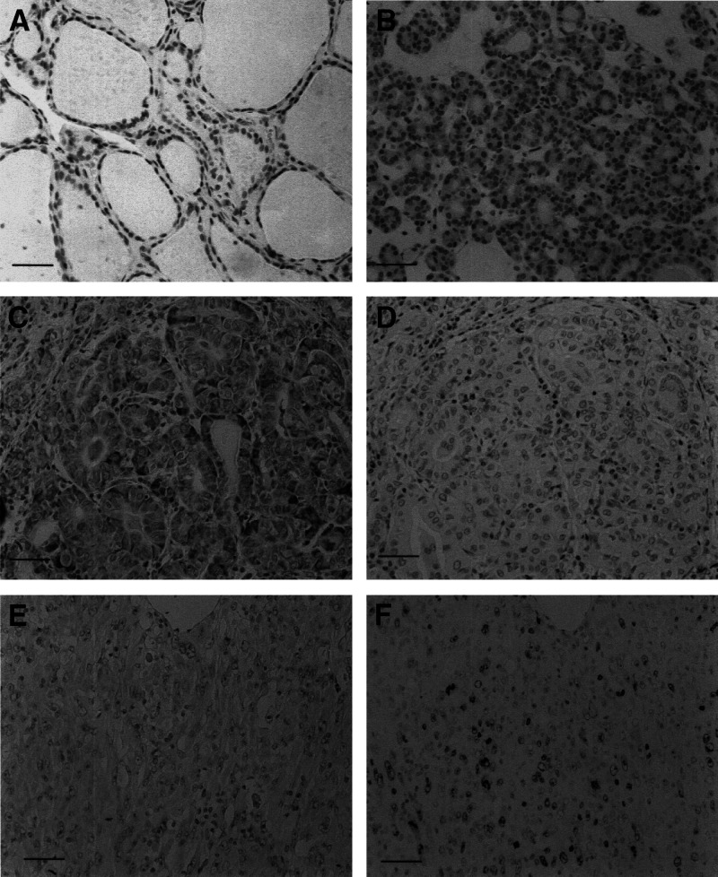
Figure 2.
Immunostaining of PLK1 and Ki-67 in thyroid tissues. (A) PLK1 was not expressed in normal follicules. (B) Absence of PLK1 expression in follicular carcinoma. (C) PLK1 overexpression in papillary carcinoma. (D) Absence of Ki-67 antigen expression in a serial section with (C). (E) PLK1 was not overexpressed in anaplastic carcinoma. (F) Ki-67 antigen expression is frequently observed in a serial section with (E) Scale bar 150 μm.

