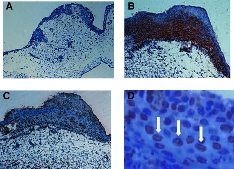
Figure 2.
Prostate cancer microtumours growing on the CAM. (A) In the mock-treated control, only very few cells of the tumour, sitting on top of the CAM, show signs of apoptosis. (B) Strong labelling (red staining) of tumour cells by immunohistochemistry with an anti-human antibody to demonstrate the human origin of the evaluated cells (MNF 116 cytoceratin cocktail (DAKO, Germany). (C) Tributyrin-treated tumours growing on the CAM. TUNEL assay reveals dark brown staining as a correlate to apoptotic changes (magnification × 20). (D) Higher magnification of (C). Nuclei-bound staining by TUNEL assay is indicated by white arrows (× 800 magnification).

