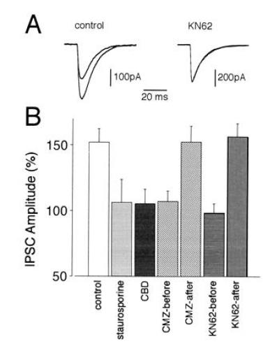
Figure 3.

Effects of staurosporine, CBD, calmidazolium, and KN62 on the rebound potentiation of spontaneous IPSCs. (A) Averaged traces (each from 50 consecutive spontaneous IPSCs) recorded in two representative Purkinje neurons from the control and KN62-treated groups, respectively. (Left) Control. Superposition of an averaged trace taken before (small amplitude) and a potentiated averaged IPSC taken 15 min after the depolarizing pulse. The averaged two traces recorded with the same protocol in the presence of KN62 (Right) are almost perfectly superimposed. Note that neither during control nor in the presence of KN62 is the time course of the IPSCs affected. (B) Average changes in IPSC amplitudes induced by the following experimental manipulations: control (n = 9), staurosporine (n = 6), CBD (n = 7), calmidazolium applied prior to and present during depolarization (CMZ-before, n = 6), calmidazolium started 5 min after depolarization (CMZ-after, n = 5), KN62 applied prior to and present during depolarization (KN62-before, n = 7), and KN62 started 5 min after depolarization (KN62-after, n = 6). Values shown represent changes of amplitudes (mean ± SEM) of 200–400 consecutive IPSCs measured 15 min after the conditioning depolarizing pulse normalized with respect to the control values before the depolarization.
