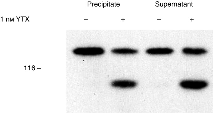
Figure 6.
Effect of yessotoxin on the distribution of E-cadherin and ECRA100 between soluble and particulate material prepared from MCF-7 cells. Cells were incubated with 1 nM YTX for 24 h at 37°C, before being processed to prepare Triton X-100 soluble (supernatant) and insoluble (precipitate) components, as described under Materials and Methods. Samples were then subjected to SDS–PAGE and immunoblotting, using the HECD-1 anti-E-cadherin antibody. The electrophoretic mobility of β-galactosidase (116 kDa) subunits, used as marker proteins running in a parallel lane, is indicated on the left.

