Figure 5.
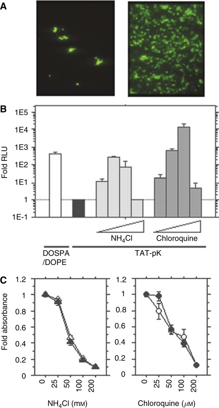
Characterisation of TAT-pK-mediated gene transfer in HEK 293 cells. (A) Enhancement of TAT-pK-mediated GFP gene expression in HEK 293 cells. Cells were seeded in 24-well tissue culture plates at a density of 2 × 10⁴ cells well−1. The cells were treated with 1 ml fresh medium containing TAT-pK/pcDNA-EGFP complex (4 μg of peptide and 1 μg of DNA) in the absence (left) or the presence of 100 μM chloroquine (right). EGFP expression was detected with fluorescent microscopy as described under Materials and Methods. (B) Comparison of transfection activity of DOSPA/DOPE/DNA complex with TAT-pK/DNA complex and effects of ammonium chloride or chloroquine on TAT-pK-mediated gene transfer. HEK 293 cells (5 × 10⁴ well−1) were seeded into 12-well tissue culture plates. The cells were treated with DOSPA/DOPE/DNA complex (2 μl of DOSPA/DOPA and 1 μg of DNA, open bar), according to the procedures recommended by the suppliers, or with TAT-pK/DNA complex (4 μg of peptide and 1 μg of DNA) as described under Materials and Methods. The cells with TAT-pK/DNA complex were incubated for 48 h in the absence (filled bar) or presence (grey bars) of ammonium chloride (25, 50, 100, and 200 mM) or chloroquine (25, 50, 100, and 200 μM). After incubation, cells were harvested and luciferase activity was evaluated. The luciferase activities were averaged from the results of duplicate experiments and are presented relative to the control value, indicated with the filled bar. (C) Cytotoxicity of ammonium chloride or chloroquine on HEK293 cells. Cells were seeded into 96-well plates and incubated at 37°C for 48 h in fresh medium containing a given concentration of ammonium chloride (left) or chloroquine (right) with (filled) or without (open) 20 μg ml−1 TAT-pK. After incubation, absorbance was measured by the WST-8 assay, as described under Materials and Methods. Each end point represents the mean ± s.d.

