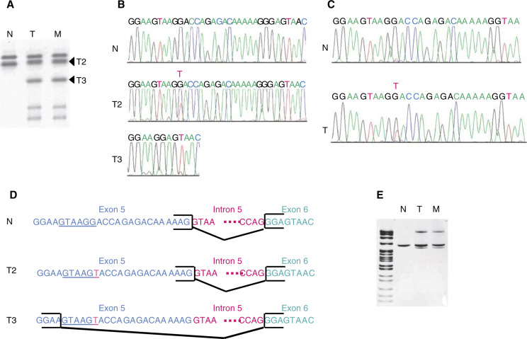
Figure 2.
PTEN mutation in the primary tumour and liver metastasis of case 26. (A) Abnormal bands were detected by SSCP analysis of cDNA from the primary tumour (T) and liver metastasis (M) using primers in exon 5 (sense) and exon 6 (antisense). These bands were not present in normal colon cDNA from the same patient (N). (B) Sequencing analysis of the two main abnormal bands (T2) and (T3) present in the primary tumour. Sequence of the normal cDNA from the same patient (N). (C) Sequencing of the genomic DNA of the primary tumour (T) and corresponding normal tissue (N). Tumour DNA harboured a G to T point mutation. (D) The various alternatively spliced forms deduced from the cDNA and genomic sequences presented in (B) and (C) are shown. The T2 allele carrying a G/T transversion in exon 5 presented the same splice form as the normal allele. T3 showed a 21 bp deletion at the 3′ end of exon 5. The new consensus donor splice site created by the mutation is underlined. (E) RT–PCR analysis of the primary tumour (T), liver metastasis (M) and corresponding normal tissue using the same primers as in (A). Lane 1: pBR322 DNA-MSPI digest.

