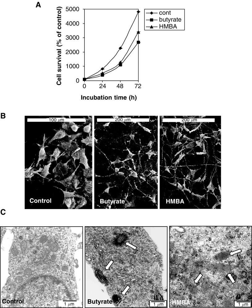
Figure 1.
Effect of butyrate and HMBA on the morphology and cell proliferation of B16 melanoma cells. The cells were incubated with 2.5 mM butyrate or 5 mM HMBA for different times. (A) Presents the proliferation rate of the cells as detected by the MTT assay. Each point is the mean ± SE of two to three separate experiments. P value <0.005. (B) Scanning electron microscopy and (C) Transmission electron microscopy. The arrows indicate the presence of melanin.

