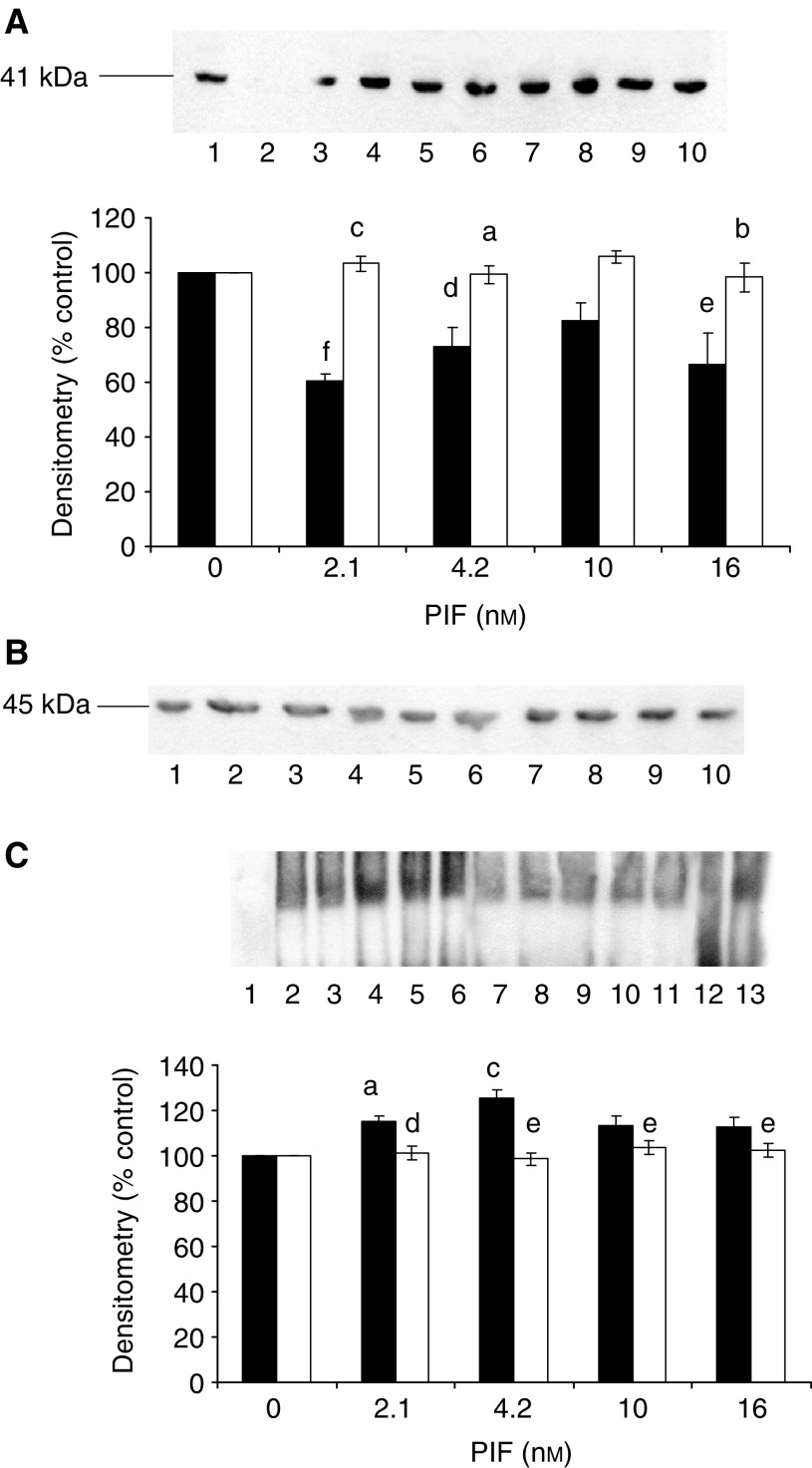
Figure 8.
Effect of PIF and calphostin C on cytoplasmic I-κBα (A) and nuclear-bound NF-κB (C) determined 30 min after PIF addition to murine myotubes. (A) Myotubes were treated with 0 (lanes 1 and 6), 2.1 (lanes 2 and 7), 4.2 (lanes 3 and 8), 10 (lanes 4 and 9) or 16.8 nM PIF (lanes 5 and 10) in the absence (lanes 1–5) or presence (lanes 6–10) of calphostin C (300 nM) added 2 h prior to PIF. (B) Actin loading control for the blot shown in (A). (C) Nuclear levels of NF-κB in myotubes in the absence (lanes 2–6) or presence (lanes 7–11) of calphostin C. Lane 1 is a negative control and lane 13 a positive control and lane 12 contains excess unlabelled NF-κB. The other lanes were the same as in (A). The densitometric analysis is an average of three replicate EMSAs. Differences from control are indicated as aP<0.05 and cP<0.001, while differences in the presence of calphostin C are indicated as dP<0.01 and eP<0.001.

