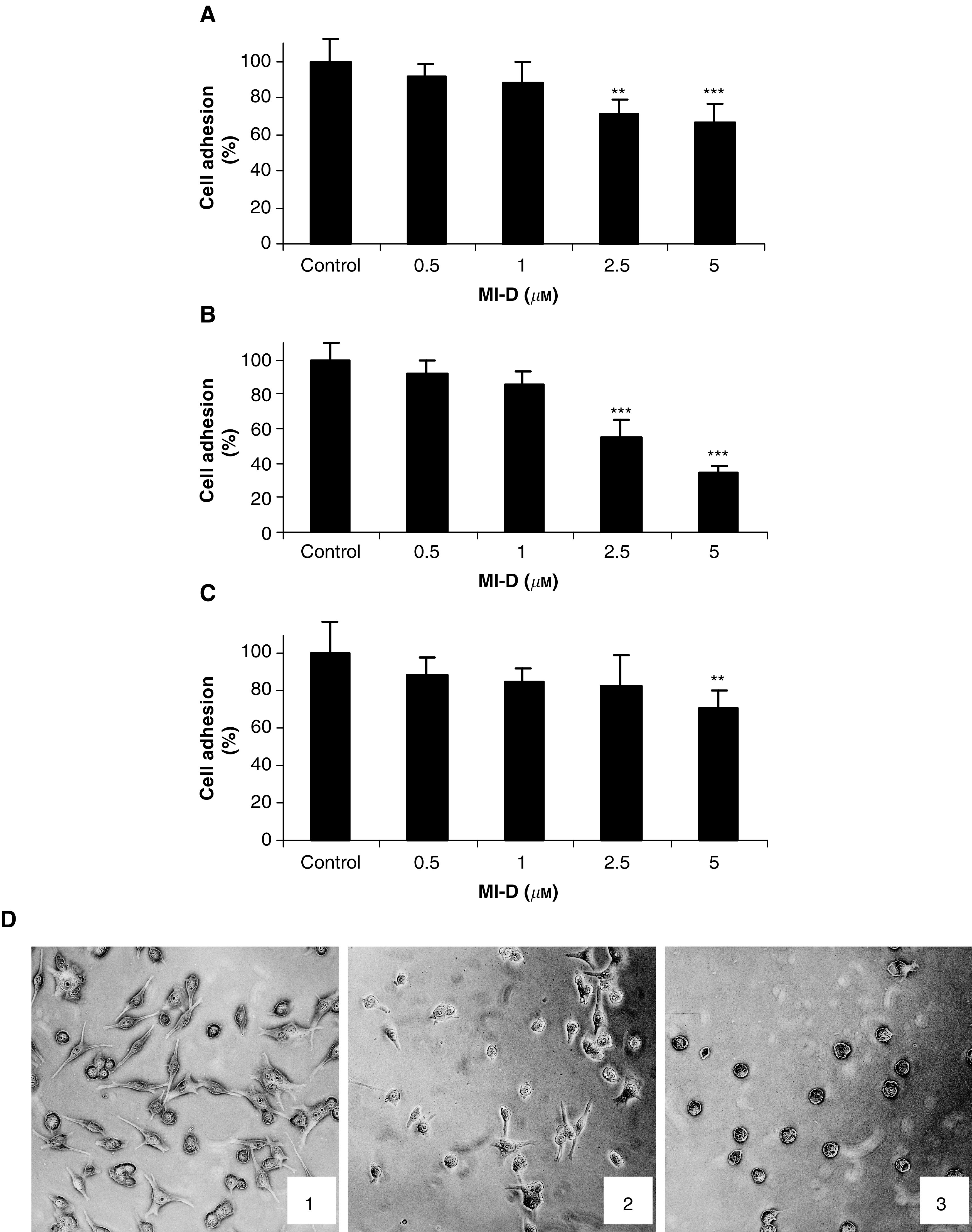
Figure 4.

Effects of MI-D on the adhesion of MEL-85 human melanoma cell to ECM constituents. (A) MEL-85 adhesion to laminin. (B) MEL-85 adhesion to fibronectin. (C) MEL-85 adhesion to matrigel. (D) Micrographs of MEL-85 cells adhered to fibronectin. (1) Control cells, (2) cells treated with 2.5 μM MI-D, (3) cells treated with 5 μM MI-D. MEL-85 cells (4 × 10⁴) were added to microculture wells precoated with ECM constituents in the presence of indicated concentrations of MI-D. After a 2 h incubation, nonadherent cells were washed and adherent cells were fixed and stained with 0.8% (p v−1) crystal violet containing 20% methanol. After extensive washing, the stained cells were lysed with 50% ethanol in 0.05 M sodium citrate and the absorbance was measured at 550 nm. **P<0.01 and ***P<0.001.
