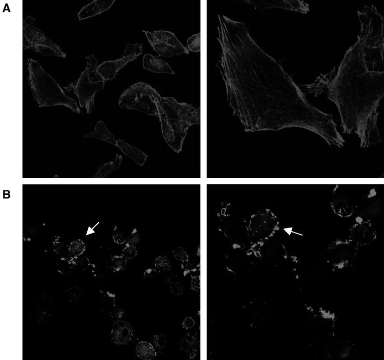
Figure 6.
Effects of MI-D on F-actin cytoskeleton organisation of MEL-85 cells. (A) Micrographs of MEL-85 control cells (× 600). (B) MEL-85 control cells at a higher magnification showing details of well-organised F-actin cytoskeleton (× 1450). (C) Micrographs of MEL-85 cells treated with 50 μM MI-D (× 600). (D) MEL-85 treated with 50 μM MI-D, cells under a higher magnification, depicting disturbances to the organisation of F-actin molecules, concentrated at the edge of cells with a granular pattern (arrow) (× 1200). MEL-85 cells were treated with MI-D, fixed and labelled with a phalloidin-FITC conjugate and observed at a confocal fluorescence microscope (Confocal Radiance 2100, Bio-Rad) coupled to a Nikon Eclipse 800 with plan apochromatic objectives.

