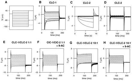
Figure 1.

Voltage-clamp traces of oocytes injected with CLC-1 cRNA (B), CLC-2 cRNA (C and D), and coinjected with both RNAs (E and F). (A) Voltage-clamp pulse protocol. Test pulses were applied in −20 mV steps starting at +40 mV (holding potential: −30 mV). The tail pulse was varied depending on the cRNA injected. Solid line, CLC-2; dashed line, CLC-1; dotted line, coinjection. (B) Typical current traces of an oocyte expressing CLC-1, showing the characteristic inward rectification at positive voltages and the deactivation at negative voltages. (C and D) Typical currents of an oocyte expressing CLC-2. Long pulses (C) elicit a slowly activating current at negative voltages, which deactivates relatively fast when stepping to +40 mV. Short pulses (as in B, E, and F) does not significantly activate CLC-2 (D). (E and F) Typical currents of an oocyte coinjected with CLC-1 and CLC-2 cRNA at a 1:1 concentration ratio. Treatment with 0.2 mM 9-AC (F) abolishes a remaining deactivating component at negative voltages, as well as the gating seen in the tail current at a potential of −140 mV. This is more obvious when oocytes injected with an about 10-fold excess of CLC-1 over CLC-2 cRNA (G) are treated with 9-AC (H).
