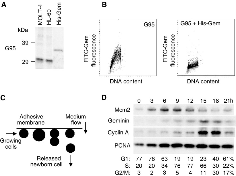
Figure 1.
(A) Immunoblots of total cell lysates prepared from asynchronously proliferating MOLT-4 and HL-60 cells and rec. human Geminin with anti-Geminin antibody G95. Rabbit polyclonal affinity-purified antibody G95 detected a single protein with a molecular mass of ∼33 kDa in total lysates from MOLT-4 (lane 1) and HL-60 (lane 2) cells, and recognised nanogram quantities of rec. Geminin (lane 3; ∼34 kDa). (B) Bivariate flow cytometry of asynchronous MOLT-4 cells with G95 alone, or G95 plus rec. Geminin, further supports the notion that the antibody is specific for human Geminin. Note that cells in G1 (2C; grey dots) are Geminin-negative, whereas cells in S–G2–M (black dots) are positive for Geminin. Pre-incubation of G95 with rec. Geminin results in loss of cell cycle periodicity and little Geminin expression is detected at any stage of the cell cycle. (C) Schematic of membrane elution. Asynchronously growing MOLT-4 cell cultures are immobilised on surfaces such that gravity coupled with cell division results in release of one daughter cell, while the other remains surface-bound. Newborn early G1-phase cells are continuously released in the effluent and grow synchronously without evidence of disturbance. (D) Immunoblots of origin-licensing factors and control proteins in synchronously proliferating MOLT-4 cells. Protein levels of Mcm2, Geminin, Cyclin A and PCNA were determined in lysates from equivalent numbers of cells isolated from synchronous batch cultures at the indicated times.

