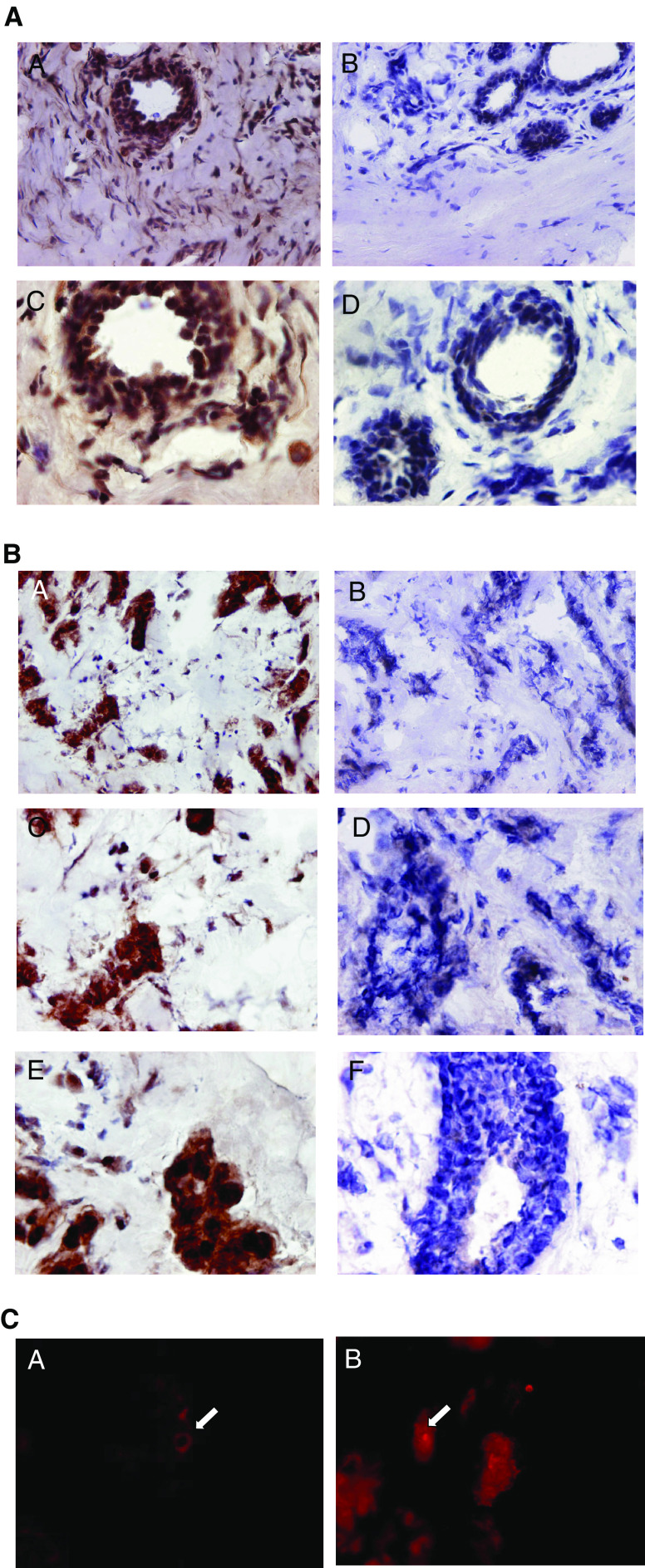
Figure 3.
(A) Ets-1 staining of frozen sections of fibroadenoma. Immunohistochemical staining of fibroadenoma tissue showing Ets-1 protein distributution mainly around the lobules and ducts and also in the stromal fibroblasts. Some negatively stained cells are also seen in the surrounding stromal tissue. Immunoperoxidase staining of sections was carried out using the Vectastain Elite ABC kit (Vector Laboratories) on 7 μm sections of frozen breast tissue embedded in OCT according to the manufacturer's recommendations. (A) Fibroadenoma tissue section × 20 (objective); (B) rabbit IgG-negative control × 20 (objective); (C) × 40 showing Ets-1 staining in the epithelial cells lining the ducts and also in the surrounding stroma; (D) IgG control × 40. (B) Ets-1 staining of frozen sections of breast tumour tissue. Immunohistochemical staining of breast tumour tissue showing Ets-1 protein distributution mainly in the tumour cells and to a lesser extent in the stromal fibroblasts surrounding the tumour islands. Some negatively stained cells are also seen in the surrounding stromal tissue. Immunoperoxidase staining of sections was carried out using the Vectastain Elite ABC kit (Vector Laboratories) on 7 μm sections of frozen breast tissue embedded in OCT according to the manufacturer's recommendations. (A) Breast tumour tissue section × 20 (objective); (B) rabbit IgG negative control × 20 (objective); (C) × 40 showing Ets-1 staining in the tumour cells; (D) IgG control × 40; (E) × 60 showing a cluster of tumour cells stained positively for Ets-1; (F) IgG control × 60. (C) Localisation of Ets-1 protein in breast tumour tissue by immunofluorescent microscopy. Ets-1 staining is observed in both the nucleus and cytoplasm of the tumour cells in the primary breast cancer tissue sections. (A) Arrow shows Ets-1 cytoplasmic staining (original magnification × 200); (B) arrow shows Ets-1 nuclear staining.

